fbpx

Click here to learn about our COVID protocols for appointments

Posts Categorized: What’s New

Eye Medical Clinic Welcomes Jeanie Paik, MD

Dr. Jeanie Paik and her family recently left windy Chicago for beautiful San Jose, and they couldn’t be happier about it! We’re thrilled that she has joined the family of eye experts at Eye Medical Clinic and are excited to introduce her to you.

Dr. Paik is a world-class physician with an elite pedigree. She earned her undergraduate degree from the University of Southern California in Biological Sciences and International Relations, and her Medical Doctorate from the University of Chicago Pritzker School of Medicine. She performed her residency at New York Eye and Ear Infirmary in New York, NY. She then completed additional training with a cornea, refractive and external disease fellowship at Emory University in Atlanta, GA.

As a board-certified, fellowship-trained specialist and surgeon, Dr. Paik brings significant experience in diseases of the cornea and anterior segment. As of this month, she will serve as our go-to expert for all corneal cases, including cornea transplants, with specialized expertise in keratoconus, corneal dystrophies, pterygium and corneal infections.

Dr. Paik performs a variety of cornea transplants, including ultra-thin DSAEK and DMEK. Dr. Paik is trained in cross-linking, an advanced treatment to help stop the progression of keratoconus. She specializes in premium cataract surgery, including multifocal and toric lenses. She is also a dry eye specialist – look for EMC to begin offering a wide range of services for dry eye soon!

While Dr. Paik performs everything from modern cataract surgery to complex cornea transplants, her favorite part of her work is the opportunity for patient connection.

Vision is so important for quality of life. I love developing relationships with my patients and caring for them over time.

Jeanie Paik, MD

Dr. Paik has presented at numerous national conferences and has published in the peer-reviewed journal Cornea. She has also published literature on allergic conjunctivitis. Her research includes topics such as keratoconus, corneal surgical techniques and corneal infections. Dr. Paik is a member of the American Academy of Ophthalmology, American Society of Cornea and Refractive Surgery and the Cornea Society.

Outside of work, Dr. Paik and her husband, a data engineer, are parents to their young daughter and enjoy spending time with family. EMC warmly welcomes Dr. Paik. We can’t wait for you to meet her. Schedule an appointment today!

Eye Medical Clinic Warmly Welcomes Tamer Hadi, MD

Tamer Hadi, MD, came to Eye Medical Clinic with an impressive resume. With undergraduate and master’s work in bioengineering and biomedical engineering, Dr. Hadi spent several years working in orthopedic research and development before deciding he wanted to be more involved in patient care. He applied to medical school and the rest is history!

With the same passion, excellence and problem-solving mindset that exemplified his earlier training, Dr. Hadi graduated with, not just a medical degree, but also a Ph.D., from the Medical College of Virginia. He didn’t necessarily enter medical school knowing he wanted to specialize in ophthalmology, but he talks about a tremendously impactful two-week rotation in ophthalmology. It was life-changing and inspiring, and he knew then it was what he wanted to do professionally.

Residency training in ophthalmology at Case Western Reserve University Medical School in Ohio led to a rigorous fellowship in vitreal-retinal surgery at the University of California in Irvine, California. Dr. Hadi joined the EMC family in August of 2020. You can see his full bio here.

I came to medicine a little later in life, but I have never regretted it. I loved my research work, but it felt a little disconnected from being able to help people in a very tangible way. All my background has now come together and allowed me to apply everything I have learned to improve the quality of life for my patients.

-Tamer Hadi, MD

At EMC, we’ve always focused on being a trusted practice that offers comprehensive eye care for patients in every stage of life. In our 70+ years, we’ve served generations of families in and around the San Jose area. We’re excited to add this powerhouse retina specialist to the team to expand our range of services even more.

Dr. Hadi truly enjoys being a doctor and a surgeon, and he especially loves interacting with his patients.

I love seeing the immediate gratitude patients experience when they discover their vision has been restored.

-Tamer Hadi, MD

Outside of work, Dr. Hadi is a husband and father of three young children. He enjoys staying active, hiking and spending time at the beach with his lively family. Together, they all enjoy working in their family vegetable garden and are expecting a bumper crop of cilantro this season!

EMC warmly welcomes Dr. Hadi. We can’t wait for you to meet him. Schedule an appointment today.

Proudly Welcoming Dr. Mona Bagga

2018 is shaping up to be a banner year at Eye Medical Clinic! Faithfully serving the San Jose area for more than 60 years, we’ve earned a solid reputation for excellence in advanced vision services, including surgery and comprehensive eye care.

We have an exciting announcement! Mona Bagga, MD, a renowned, board-certified ophthalmologist with fellowship training in cataracts and glaucoma, will join our team in early 2018. This new partnership takes our practice to the next level!

“I’m excited to join Dr. Sullivan and the amazing team at Eye Medical Clinic. I’m ready to help bring the practice to the next level with innovative procedures for glaucoma and cataracts,” said Dr. Bagga. “Even though the practice has such a great history in San Jose, in many ways I feel like it’s a newborn that I get to help raise. I’m excited to bring pioneering procedures to restore sight and improve lives, and I can’t wait to delight our patients.”

Dr. Bagga brings a lot to the table, ophthalmologically speaking (say that 10x fast!). She has deep experience in cataract and glaucoma treatment, and uses a combination of advanced medical, surgical, and laser procedures to improve her patients’ vision and quality of life. In addition to thousands of conventional glaucoma surgeries, such as trabeculectomy and glaucoma drainage implants, Dr. Bagga has performed hundreds of minimally-invasive glaucoma procedures using iStent, Trabectome, and Canaloplasty. She was the first surgeon in Arizona to perform the iStent procedure for glaucoma.

Dr. Bagga’s skill set is a welcome addition, and we are very excited to add cataract surgery, premium lens implants, and the surgical treatment of glaucoma to the list of services we are now able to offer our patients.

Since 1996, Dr. Bagga has completed over 10,000 cataract surgeries, including laser-assisted cataract surgery, multifocal, and toric implants. She was one of the first two surgeons in the United States to implant the Tecnis Toric Lens! Dr. Bagga is actively involved in clinical research and has authored numerous publications, lectured nationally and internationally, and served as an investigator in numerous clinical trials.

Dr. Bagga is very well respected in her field and is a highly sought-after surgeon. “I am very excited to move to the San Jose area as I have a lot of family and friends there,” said Dr. Bagga. “This is where I want to be. My vision matches very well with the vision of Eye Medical Clinic. There is a lot of potential for growth and I can’t wait to jump in.”

Dr. Bagga may love surgery the most, but in her spare time, she enjoys painting, as it appeals to her artistic nature. She also enjoys hiking and spending time with family and friends.

The entire Eye Medical Clinic team warmly welcomes Dr. Bagga!

Ultraviolet Light and your Eyes

Ultraviolet Light and your Eyes

If you want strong, healthy eyes and clear vision for life, a major step you can take is to protect your eyes from UV radiation.  Wearing proper eye protection from the sun reduces the risk of a number of eye diseases and other conditions that are caused or worsened by UV exposure.

Eye Diseases Linked to UV Exposure

UV exposure has been linked to a number of serious eye diseases including macular degeneration and cataracts.

Macular Degeneration

Macular degeneration is a condition in which the macula of the eye breaks down, leading to a loss of central vision and is a leading cause of age-related vision loss.  Macular degeneration develops over time so a lifetime of exposure to UV can contribute it’s likelihood.

Cataracts

Cataracts occur when the natural lens of the eye becomes clouded, resulting in blurred vision and eventually blindness. The len is responsible for focusing the light that comes into the eye, allowing clear vision. Cataracts can be treated by a simple surgery to replace the clouded lens with an artificial lens.  UV light contributes to certain types of cataracts, which account for about 10% of all cases.

Skin Cancer

Another serious disease that can affect the eyes is skin cancer which can appear on the eyelids or the area around the eyes.  Skin cancer is known to be linked to extended exposure to UV and your eyes can be a difficult area to protect with sun block as you don’t want it to get too close to the eyes.

Other Eye Conditions Linked to UV Exposure

Photokeratitis or Corneal Sunburn

Photokeratitis or a corneal sunburn in layman’s terms can occur with intense exposure to the sun without proper eye protection. It is commonly experienced after a day skiing or snowboarding at a high altitude or at the beach. Corneal sunburns can be extremely painful and can sometimes cause a temporary loss of vision.

Pterygium

Pterygium, also known as “surfer’s eye” is a growth that forms on the conjunctiva which is a layer over the sclera or the white part of your eye. Sometimes they grow onto the cornea as well. Often pterygia are harmless but if they grow too large they may begin to impact your vision. In this case, surgery may be necessary. Pterygia are commonly found in individuals who spend a significant amount of time outside in the sun or wind.

How to Properly Protect Your Eyes From UV

The more time you spend outside, the greater the risk for your eyes, however you can easily minimize this risk with proper protection. Here are a few tips to ensure you are doing what you can to safeguard your eyes:

Proper Sunglasses

Fully protective sunglasses should block out 99-100% of UV-A and UV-B rays. You can achieve this through purchasing a pair of sunglasses, applying a UV blocking coating to your glasses or opting for photochromic lenses which are eyeglass lenses which turn dark when exposed to sunlight. Most contact lenses will also have UV protection but this is just for the area of the eye covered by the lens.

Since UV exposure can enter from the air, the ground or from the sides, wrap-around and large lensed frames can provide added protection.

Add a Wide Brimmed Hat

A wide brimmed hat or visor will stop about half of the UV rays from even reaching your eyes as well as reduce the exposure coming in from the top or sides of your sunglass frames.

Know Your Environmental Risk Factors

UV exposure is largely dependent upon your location and your surroundings. If you are located at a high altitude you will likely be exposed to more UV than at lower altitudes.  UV also reflects off of snow, sand, water and even asphalt so be aware that you are getting increased exposure under these conditions.

Know Your Additional Risk Factors

There are a number of other factors that can increase your exposure or risk of eye damage from UV.  For example, certain medications increase the sensitivity of your eyes and skin to sunlight (speak to your doctor about any medications you are on). Previous eye surgery or eye diseases can also increase your risk factors for UV eye damage. Additionally if you work in certain fields such as welding or medical scans or radiation or use tanning beds, you can be exposed to additional UV radiation. If there is nothing you can do to change your exposure, make sure you are properly protecting your eyes with goggles or glasses and a hat.

Regular Eye Exams

Make sure you schedule a comprehensive eye exam on a regular basis to ensure your eyes are healthy.  If you are over 50 or have increased risk factors for eye disease, you should schedule exams at least on a yearly basis or according to your eye doctor’s recommendations.


Signs of Eye and Vision Problems in Infants

Signs of Eye and Vision Problems in Infants

Infant Eyesight

Despite nine months of growth in utero, babies are not born with fully developed eyes and vision – just like they can’t walk or talk yet. Over the first few months of life, their visual systems continue to progress, stimulated by their surroundings.

Babies will develop the ability to track objects, focus their eyes, and move them like a team. Their visual acuity will improve and they will gradually be able to see more colors. They will also form the neural connections that will allow them to process what they see, to understand and interact with the world around them.

Healthy eyes and good vision are necessary for proper and timely progress; ocular or visual problems can lead to developmental delays.

So how do you know if your infant is developing normally? What can you do to ensure your baby’s eye health and vision are on track? While infant eye problems are not common here are some steps you can take to ensure your child’s eyes are healthy.

#1 Schedule a six month check-up.

It is recommended to get the first professional comprehensive eye and vision exam for your child between six and 12 months of age.

Your optometrist should check for the following skills at the 6-month checkup:

  • Visual acuity (nearsightedness, farsightedness or astigmatism)
  • Eye muscle and movement capabilities
  • Eye health

If you have any concerns prior to six months, don’t hesitate to take your baby for an exam earlier.

#2 Engage in visually stimulating play.

Incorporating visually stimulating play for your child will help develop visual processes like eye tracking and eye teaming.

A baby’s initial focusing distance is 20-30 cm, so to nurture healthy vision skills, keep high contrast “reach and touch” toys within this distance. Alternate right and left sides with each feeding, and provide toys that encourage tracking of moving objects to foster eye-hand coordination and depth perception.

Pediatricians in North America recommend that NO screen time be allowed under the age of 2, as many forms of development may be delayed from premature use of digital devices.

#3 Be alert to eye and vision problems.

Keep an eye out for indications of an eye health problem, and contact an eye doctor to discuss any concerns you may have. Some symptoms to pay attention to include:

  • Red eyes or eyelids, which may or may not be accompanied by discharge and crusty lids. This may indicate an eye infection that can be very contagious and may require medication.
  • Excessive eye watering or tearing. This may be caused by a problem with the tear ducts, such as a blockage.
  • Extreme light sensitivity. While some light sensitivity is normal, significant sensitivity to light can be a sign of disease or elevated eye pressure.
  • Eye “jiggling” or bouncing. This suggests a problem with the muscle control of the eyes.
  • Eye turn. Whether it is an eye that seems to cross in or a “lazy eye” that turns out, this is often associated with a refractive error or eye muscle issues that could require treatment such as eyeglasses, vision therapy, patching or surgery.
  • White pupil. This can be a sign of a number of diseases, including cancer. If you see this have it checked out immediately.

Since your infant’s eyes are still maturing, any issues that are found can likely be corrected with proper care and treatment. The important thing is to find a pediatric eye care provider that you trust because you will want to regularly check the health of your child’s eyes to ensure proper learning and development throughout infancy and beyond.

Trouble Seeing at Night? All About Night Blindness


At this time of year when the sun sets early, many people are affected by night blindness. Night blindness or nyctalopia refers to difficulty seeing at night or in poor or dim lighting situations. It can be caused by a number of underlying conditions, sometimes completely benign and sometimes as a symptom of a more serious eye disease. So, if you are experiencing trouble seeing in low light, especially if it is a sudden onset of the condition, it is worth having it checked out by your eye doctor.

Signs of Night Blindness

The main indication of night blindness is difficulty seeing well in dark or dim lighting, especially when transitioning from a brighter to a lower light environment, like walking from outside into a dimly lit room. Many experience difficulty driving at night, particularly with the glare of the streetlights or the headlights from oncoming traffic.

Causes of Night Blindness

Night blindness is a condition that can be present from birth, or caused by a disease, injury or even a vitamin deficiency. In order to treat the condition, your eye doctor will need to determine the cause. Here are some of the common causes:

  • Nearsightedness (myopia) – many people with nearsightedness (or difficulty seeing objects in the distance) experience some degree of night blindness, especially when driving.
  • Retinitis Pigmentosa – a genetic condition in which the pigmented cells in the retina break down causing a loss of peripheral vision and night blindness.
  • Cataracts – a clouding of the natural lens of the eye causing vision loss.
  • Glaucoma – a group of diseases that involve damage to the optic nerve and subsequent vision loss.
  • Vitamin A Deficiency – vitamin A or retinol is found in greens (kale, spinach, collards, broccoli etc.), eggs, liver, orange vegetables (carrots, sweet potatoes, mango etc.), eggs and butter. Your doctor may also prescribe Vitamin A supplements if you have a serious deficiency.
  • Eye Surgery – refractive surgery such as LASIK sometimes results in reduced night vision as either a temporary or sometimes a permanent side effect.
  • Injury – an injury to the eye or the part of the brain that processes vision can result in reduced night vision.
  • Uncorrected Visual Error – many people experience better daytime vision as the pupils are smaller and provide greater depth of field to compensate for any vision problems. At night, the pupils dilate, so blur is increased from uncorrected nearsightedness, farsightedness, astigmatism or distortions/aberrations on the cornea from refractive surgery. Even a slight prescription for someone who may not need glasses during the day can make a significant improvement in night vision.
  • Eyewear Problems – even if your vision correction is accurate, badly scratched glasses or poor/defective lens coatings can also cause trouble seeing at night. Special lens coatings are now available on glasses for night time and foggy conditions.

Treatment for Night Blindness

Some causes for night blindness are treatable, while others are not, so the first step is a comprehensive eye exam to determine what the root of the problem is. Treatments range from simply purchasing a special pair of glasses, lens coatings or contact lenses to wear at night (for optical issues such as myopia) to surgery (to correct the underlying problem such as cataracts), to medication (for diseases like glaucoma). In some cases, your doctor may recommend that you avoid driving at night. During the day, it may help to wear sunglasses or a brimmed hat to ease the transition indoors.

As with any change in vision, it is critical to get your eyes checked as soon as you begin to experience symptoms, and on a routine basis even if you’re symptom-free. Not only will this improve your chances of detecting and treating a vision-threatening disease if you have one brewing, but treatment will also keep you more comfortable seeing in low-light, and keep you and your loved ones safe at night or in poor light conditions.

Are Your Eyes Sensitive to Light?


Light sensitivity, also known as photophobia, is a condition in which bright light – either natural sunlight or artificial light –  can cause significant discomfort, pain and intolerance. People that experience light sensitivity will find themselves needing to close their eyes or squint when exposed to light and often experience headaches and nausea as well.  In mild cases, the discomfort accompanies exposure to bright lights or harsh sunlight, but in severe cases even a small amount of light can cause pain and discomfort.

Photophobia is more common in individuals with light eyes. This is because the greater amounts of pigment in darker eyes help to protect the eye from the harsh rays of light. The darker pigment of the iris and choroid absorbs the light, rather than reflecting the light and causing internal reflection or glare experienced by those with lighter eyes. People with albinism, which is a total lack of eye pigment, also experience significant light sensitivity for this reason.

Acute photophobia is usually a symptom that accompanies a condition such as an eye infection or irritation (such as conjunctivitis or dry eyes), a virus, or a migraine (light sensitivity is one of the most common symptoms of migraines). It could also be caused by something more serious such as an eye condition like a corneal abrasion, a detached retina, uveitis or iritis or a systemic disease like meningitis or encephalitis. Light sensitivity is also a side effect of refractive surgery (such as LASIK) and some medications (such as tetracycline and doxycycline).

How to Deal with Photophobia

The most effective way to reduce the discomfort caused by photophobia is to stay out of sunlight and dim indoor lights as much as possible while you are experiencing symptoms. Wearing dark sunglasses and keeping your eyes closed may also provide some relief.

In the summer it is more common for UV to trigger corneal inflammation (keratitis) and cause photosensitivity as well. Wind and eye dryness can also set off photosensitivity, which are more good reasons to wear sunglasses.

If the sensitivity is new and the cause is unknown, you should seek medical attention immediately, especially if you experience any of the following symptoms:

  • Blurry vision
  • Burning or pain in the eye
  • Fever and chills
  • Confusion and irritability
  • Severe headache
  • Drowsiness
  • Stiff neck
  • Nausea and vomiting
  • Numbness
  • Foreign body sensation

In cases where the photophobia is a symptom of an underlying issue, treating the issue will likely cause relief in your sensitivity. This will vary depending on the ailment but could include pain medications, eye drops or antibiotics, or anti-inflammatory medications. If the sensitivity is mild due to your genetic predisposition or a result of surgery, make sure you take your sunglasses every time you leave the house. People who wear prescription eyeglasses may consider photochromic lenses which automatically darken when exposed to light.

If you are uncomfortable, speak to your eye doctor about the best options for your condition.

Got a Shiner!


What Exactly Is a Black Eye?

A black eye, also known as a periorbital hematoma, is usually not an injury of the actual eye (which is why it is called “periorbital”- around the eye). It typically occurs when there is an injury to the face or the eye socket which causes bleeding beneath the skin and bruising. The term, “black eye” comes from the dark coloring of the bruising that occurs underneath the skin around the eye.

When a blunt force hits the eye socket, this can cause capillaries in the area to burst, causing hemorrhaging, also known as a hematoma. This blood can accumulate in the eye socket and as it begins to be reabsorbed into the surrounding tissues, the colors of the bruising begin to change. That’s why you will often notice the coloring of the black eye to go from a dark purplish-red color to brownish and then yellow.

Sometimes along with the external bruising, you might also notice a small amount of bleeding on the white surface of the eye, which is called a subconjunctival hemorrhage. This is when the tiny blood vessels on the white sclera are broken and leak blood. It’s generally harmless but sometimes looks scarier to the patient than the black eye does. This condition will also reabsorb on its own and is nothing to be concerned about.

While most black eyes can look pretty serious due to the dramatic color, an uncomplicated black eye will typically heal within a week to ten days. If it doesn’t, there could be a more serious issue such as a bone fracture or an orbital blowout fracture. This could present with restricted eye movement, especially if looking up or down, and numbness of the cheek and/or upper lip on the same side as the black eye. The eye may even appear sunken in. Further, if there is bleeding within the actual eye (called a hyphema) or floaters or flashes in the vision, then it is definitely advisable to see your eye doctor as soon as possible. These could be signs of more serious damage such a corneal or retinal damage and can lead to vision loss.

Treatment for a Black Eye

Usually, the best treatment for a black eye is to apply a cold compress (or even better, a bag of frozen vegetables, which is more flexible and can conform to the contours of the face) directly on the area. The cold will reduce swelling and constrict capillaries to reduce internal bleeding as well. Apply the cold for about 15-20 minutes every hour.  If there is pain, over the counter pain medications can help.

If however, you experience any of the following symptoms, you should seek medical attention:

  • Blood on the surface of the eye or a visible incision on the eye
  • Vision changes such as double vision, blurred vision, loss of vision or the appearance of floaters
  • Loss of consciousness, dizziness or fainting
  • Loss of eye movement
  • Persistent headaches
  • Blood or fluids coming from the ears or nose
  • Vomiting
  • Signs of infection such as excessive swelling, pus, redness or a fever
  • Severe pain

In addition to blunt trauma, black eyes can be caused by sinus infections, nasal or eye surgery or other infections in the area such as the teeth infections or cellulitis (a serious infection that can occur around the eyes). A skull fracture can also cause both eyes to turn black, sometimes known as raccoon eyes.

Unless you notice any severe symptoms you can rest assured that your black eye is a bruise just like anywhere else on the body and with a little care, rest and patience, it will clear up in no time.

Cataracts: Everything You Need To Know

Everything You Ever Needed To Know About Cataracts

According to the American Academy of Ophthalmology (AAO), cataracts affect more than 24.4 million Americans age 40 and older. And by age 75, approximately half of all Americans have cataracts. As the U.S. population ages, more than 30 million Americans are expected to have cataracts by the year 2020.

The good news is that cataracts are very treatable. More than 2 million men and women undergo cataract surgery every year, making the procedure one of the most common and successful medical procedures performed in the U.S. today.

What Is A Cataract?

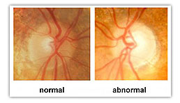
A cataract is a clouding of the usually transparent lens in your eye. The lens focuses light onto the retina at the back of your eye, allowing you to see. When your natural lens starts to cloud up, the images projected onto your retina become blurry and unfocused. It’s like looking through a dirty or cloudy window. If the window is not clear, you can’t see!

Cataracts are a natural part of the aging process, so if you live long enough, you will likely eventually develop one.

Symptoms Of Cataracts

Cataracts come with warning signs; they don’t suddenly develop overnight. Symptoms vary from person to person but common symptoms include:

“Washed Out” Vision or Double Vision

This occurs when people and objects appear hazy, blurred or “washed out” with less definition, depth and color. This makes many normal daily activities, including reading, watching television, driving or doing basic chores, a challenge.

Increased Glare Sensitivity

This can happen both from outdoor sunlight or light reflected off shiny objects indoors. Glare sensitivity can cause problems with driving, particularly at night.

Dulled Colors

This is when colors don’t appear as vibrant as they once did. Distinguishing colors may become difficult as well.

Compromised Contrast And Depth Perception

These important eye skills are greatly affected by the damage to the lens a cataract can cause.

Darkened Vision

This often affects individuals with cataracts, causing them to need more light than they used to in order to see clearly and perform basic activities, especially reading.

(Insert Cataract Self Evaluation Quiz)

Click here to take our cataract self-evaluation quiz and discover if it’s time for your cataract screening.

Causes Of Cataracts (Risks)

Age is not the only risk factor for cataract development although it is the primary one. While the risk of developing a cataract does increase as you age, it is not the only factor. Other risk factors include:

  • Family history
  • Injury to the eye
  • Certain medications (e.g., steroids)
  • Diseases such as diabetes and macular degeneration
  • Alcohol consumption
  • Smoking
  • Prolonged exposure to the sun
  • Certain eye conditions such as uveitis
  • Cataracts can also be congenital (present at birth)

What You Can Do To Reduce Your Risks

Don’t let cataracts interfere with your quality of life. The best preventive measure is being sure to schedule regular eye exams so that you stay on top of your overall eye health.

While doctors still don’t know exactly how much each risk factor leads to cataracts, there are a few ways you can keep your eyes healthy and reduce your risk of developing a cataract:

  • Refrain from smoking and high alcohol consumption
  • Exercise and eat well, including lots of fruits and vegetables that contain antioxidants
  • Protect your eyes from UV radiation from sunlight
  • Control diabetes and hypertension
  • Most importantly, see your eye doctor regularly for a comprehensive eye exam. If you are over 40 or at risk, make sure to schedule a yearly comprehensive eye exam.

Because cataracts typically develop slowly over a long period of time, most people don’t even realize the lens in the eye is affected until their vision becomes dull or blurry. When a cataract begins to affect your ability to perform normal daily tasks, it is time for treatment.

Cataracts cannot be treated with medication, dietary changes or eye drops. They will not heal or even improve on their own. Surgery is the only option. Fortunately, modern cataract surgery is low risk and enjoys an excellent success rate. It is relatively non-invasive, often requiring no more than a tiny laser-assisted incision, performed in an outpatient setting.

Eye Medical Clinic is a regional leader in modern cataract surgery. Our own Dr. Mona Bagga is a board-certified ophthalmologist with fellowship training in cataract surgery. She is experienced, having completed more than 10,000 cataract surgeries in her career (so far!) and is a respected regional leader in modern cataract surgery.

Cataract Surgery

Cataract surgery is one of the most common surgeries performed today.
It is regarded as one of the safest and most effective surgeries performed in America, having a 90% success rate (patient has improved vision, between 20/20 and 20/40 following the procedure).

Traditional versus Laser-Assisted Cataract Surgery

The most recent breakthrough in cataract surgery is the introduction of the cataract laser. Eye Medical Clinic is always at the front of the line where new technology is concerned, offering the latest FDA-approved laser cataract technology for your cataract surgery procedure. The experienced, board-certified surgeons believe this technology adds to the safety, accuracy and precision of the procedure.
Laser cataract surgery, like traditional cataract surgery, is a safe procedure in which your eye’s cloudy natural lens is gently removed and replaced with a clear artificial lens implant. But unlike traditional bladed cataract surgery where the incisions are made by hand, the laser automates key steps of the cataract surgery procedure.
Its software system analyzes high-resolution images of your eye, helping your surgeon design and perform a highly customized procedure. To further enhance accuracy, a patient interface connects your eye to the image-guided surgical unit, so both the computer, and the surgeon operating it, have precise, real-time images throughout the entire procedure.

Intraocular Lenses (IOLs)

A monofocal lens is not considered a Premium Lens. It is the basic lens that would be covered by Medicare and private insurance. Although standard cataract surgery has been proven very successful at removing cataracts and replacing the eye’s natural lens with a clear monofocal lens, it is important to know that many people find they still need glasses, bifocals or multifocals in order to achieve their best vision.

Compared to standard intraocular lenses, Premium Lens Implants can greatly reduce or even eliminate the need for glasses after your cataract surgery procedure. It is important to note, however, that unlike traditional cataract technology and surgery options, Premium Lens Implant packages are not covered entirely by Medicare and insurance payers.

Premium Lens Implants

Major advances in lens technology allow people just like you to continue to live your life the way you want to live it – even after cataract surgery. Eye Medical Clinic offers a variety of Premium Lens Implants that can correct your cataracts AND get you out of your glasses or bifocals.

Multifocal Lenses are created to improve distance and near focus, while Toric lenses are designed to correct astigmatism. While there are no lens options that can definitively promise 20/20 vision at all distances, most people who choose a Premium Lens Implant for their cataract surgery procedure at Eye Medical Clinic are glasses-free for most daily activities.

Before the Surgery

Prior to cataract surgery at Eye Medical Clinic, you will have a complete eye examination that includes a thorough medical history. This exam will be performed by one of our experienced board-certified ophthalmologists.

You’ll also have a pre-operative consultation that will include a discussion about your expectations for your vision following cataract surgery. It’s very helpful to come with a list of questions and concerns so that you leave the consultation feeling confident and fully informed about your surgery.

During the Surgery

During your cataract surgery, your Eye Medical Clinic surgeon will make a tiny incision in your eye to remove the cloudy lens and replace it with a clear artificial lens. The new lens can’t be seen or felt and is designed to stay inside the eye permanently. The incision is so small that no stitches are required and it will heal completely on its own. No hospital stay required!
Post-Surgery

Thanks to advances in technology, recovery from cataract surgery is remarkably fast. Most people return to normal activities within 24 to 48 hours of their procedure but strenuous physical activity should be avoided for several weeks. You will not be able to drive home from the procedure and shouldn’t get behind the wheel until you have been given approval by your doctor after a follow-up exam the next day.

The eye may take several weeks to heal completely – your Eye Medical Clinic doctor will guide you through the postoperative process for best results.

Restored Vision, Renewed Outlook

Eye Medical Clinic is proud to offer the latest techniques and technologies in the field of cataract surgery. If you’re ready to take the next steps toward enjoying restored vision and getting back to what it is you like to do with your life, schedule your cataract evaluation today.

Are You Ignoring Your Dry Eyes?


You Don’t Have to Live With Dry Eyes

Have you noticed that your eyes feel chronically dry, itchy, scratchy or even sometimes watery? Many people that have these symptoms just go on with their lives until the symptoms become unbearable. What they don’t realize is that these are signs that they might be suffering from dry eye syndrome, a condition in which the eyes are not able to produce enough tears to effectively lubricate the eyes. This is a problem that won’t just go away on its own.

What causes Dry Eye?

Dry Eye Syndrome, also known as Tear Film Dysfunction is characterized by a reduction in the amount or quality of tears that are produced. Tears are essential for optimal eye health, vision and comfort. Ideally, tear film covers the eyes at all times to prevent the eyes from drying out and to ensure clear vision. If the glands that produce tears start to produce fewer tears or tears that don’t have the proper balance of water, oils, proteins and electrolytes, the tear film will become unstable, allowing dry spots to form on the surface of the eye, and cause disruptions in outer barrier of the eye’s epithelial surface. This disruption in the outer barrier allows microbes to invade the eye, leading to irritation and infection. The condition can be caused by many factors, including tear gland dysfunction, eyelid problems, medications or environmental factors.

Symptoms of Dry Eye

As mentioned above, many of the symptoms of dry eye involve varying sensations of dryness including, burning, stinging, itching, grittiness, soreness or a feeling that there is something in the eye. The eyes may also be red and sensitive to light, wind or smoke. Vision may be blurred or doubled and the eyes may fatigue easily. Another common symptom is that vision seems blurry but clears when you blink (especially common when reading or using a computer). This is because the tear film does not form a smooth coat over the eye surface or it evaporates too quickly causing a blur.

You may also notice pain, some discharge from the eye (especially upon waking in the morning) and experience discomfort when wearing contact lenses. One of the most confusing symptoms of dry eye is actually excessive tearing, which occurs because the eyes are trying to compensate for the lack of moisture – however the tears produced are low quality and don’t properly hydrate the surface of the eye.

Reducing Symptoms

The first thing to look at when you have dry eyes is whether you are taking any medications, engaging in certain behaviors or being exposed to environmental factors that may be causing the condition. Medications that may cause dry eye as a side effect include:

  • Antihistamines and Decongestants
  • Diuretics
  • Sleeping pills
  • Birth Control pills
  • Antidepressants
  • Acne medications
  • Angiotensin-converting enzyme (ACE) inhibitors
  • Opiate-based painkillers such as morphine

Important! Never stop medication without the approval of your doctor! If you are taking a medication that may be causing dry eye, don’t stop taking the medication without speaking to your healthcare provider first. Treating dry eye symptoms may be a simpler solution than stopping or switching medications.

You may be able to alter your environment to reduce symptoms of dry eye as well. Environmental factors that can exacerbate dry eye include:

  • Wearing contact lenses
  • Extended use of computers or other electronic devices
  • Exposure to dry, windy climates or blowing air (such as an air conditioner or heater).
  • Exposure to smoke
  • High altitudes

Treatment for Dry Eye

If you are experiencing dry eye symptoms, make an appointment with your optometrist. The diagnosis and treatment will be based on a complete examination of your eyes, your medical history and your personal circumstances around the condition. The doctor may decide to perform a tear film test that can determine the quantity and quality of the tears and whether your tear glands and tear film are functioning properly.

The type of treatment will depend on the underlying cause of the problem. Treatment may include behavioral or environmental changes such as using a humidifier, wearing sunglasses in windy weather, reducing computer time or changing to a different type of contact lens, as well as medical treatments that may include:

  • Artificial tears, eye drops or ointments to lubricate eyes
  • Steroid or antibiotic drops or pills may be used for certain conditions such as blepharitis
  • Reducing the drainage of tears by blocking tear ducts with silicone plugs
  • Medications such as Restasis which reduce inflammation and trigger tear production
  • In some situations a surgical procedure might be recommended
  • Scleral lenses that hold moisture on the surface of the eyeball

The most important thing you should know about dry eyes is that you do not have to suffer. Treatments are available to increase moisture on your eye and reduce the uncomfortable and sometime debilitating symptoms. If you are suffering, schedule an appointment with your eye doctor and get the relief you deserve.

April is Women’s Eye Health and Safety Month


Hey women! Did you know that women are more likely to suffer from vision problems and are at higher risk of permanent vision loss than men? Well 91% of the women surveyed recently didn’t know that, which means that many of them aren’t taking the necessary precautions to prevent eye damage and vision loss.

According to a recent study, the statistics for many of the major vision problems show that women have a higher percentage of incidence than men. These include:

  • Age-related Macular Degeneration 65%
  • Cataracts 61%
  • Glaucoma 61%
  • Refractive Error 56%
  • Vision Impairment 63%

Women are also more susceptible to develop chronic dry eye, partially because it is often associated with other health issues that are more common in women such as ocular rosacea which is three times more prevalent in women.  Hormonal changes during pregnancy and menopause can also contribute to dry eye.

It’s important for women to know the risks for eye-related diseases and vision impairment and the steps they can take to prevent eventual vision loss.  Here are some ways that you can help to protect your eyes and save your eyesight:

  • Find out about family history of eye diseases and conditions.
  • Protect your eyes from the sun by wearing 100% UV blocking sunglasses when outdoors.
  • Don’t smoke.
  • Consume a healthy diet with proper nutrition and special eye health supplements as prescribed by an eye doctor.
  • Adhere to contact lens hygiene and safety.
  • Adhere to cosmetic hygiene and safety precautions.
  • Protect your eyes against extended exposure to blue light from computers, smartphones and LED lamps.
  • If you are pregnant or planning to become pregnant and have diabetes, see an eye doctor for a comprehensive eye exam. In women who have diabetes, diabetic retinopathy can accelerate quickly during pregnancy and can present a risk for the baby as well.

Mothers are often charged with caring for the eye health of the entire family, but too often their own eye health needs fall to the wayside. It is critical that mothers take care of their eyes and overall health so that they can be in the best condition to care for their families.

Speak to your eye care professional about your personal eye health and vision risks and the precautions and measures you should take to protect your eyes.  Encourage the other women in your life to do so as well.  Once vision is lost, it often can’t be regained and there are many steps you can take to prevent it with proper knowledge and awareness.

The most important way to prevent vision loss is to ensure you schedule regular eye exams. Don’t wait for symptoms to appear as many eye issues are painless and symptomless, and sometimes by the time you notice symptoms, vision loss is untreatable.

It’s Time to Talk About Blue Light

Workplace Eye Safety Month

Blue light. Do you know what it is? Do you know where it comes from, or how it can be harmful to your eyes? If you don’t know the answers to these questions, you are not alone, yet it is important that you become aware to protect your eyes for now and the years to come.

The reason blue light is suddenly becoming a big issue is because other than the sun, which is the biggest source of blue light, a significant source of blue light emission comes from digital devices and artificial lighting. As our world becomes increasingly digital – think: HD televisions, LED lights, computers, smartphones, tablets – we are all exposing our eyes to more and more amounts of blue light than ever before. And we are only beginning to understand the long term effects this has on our bodies and our eyes.

One of the biggest issues with blue light is that whether it is through work or leisure, people are exposed to screens at a close range for a large portion of the day. A survey from the Vision Council entitled, “Blue Light Exposure and Digital Eye Strain” recently showed that 87% of respondents used digital devices for more than two hours a day and over 52% regularly used two digital devices as the same time. This shift has drastically increased exposure and the number of symptoms that are reported. To date, research has shown that there are a number of ways blue light can impact your eyes including digital eye strain, sleep disturbances and retina damage that can lead to long term problems including serious eye diseases.

Digital eye strain is a condition that is characterized by dry, sore, tired or burning eyes, eye fatigue and sensitivity to light. It can also cause blurred or double vision, headaches, back, neck and shoulder aches and difficulty focusing or concentrating. These symptoms are most common in individuals that sit in front of the computer for two or more hours a day.

Studies show that exposure to blue light right before bedtime can cause disruptions in sleep and wakefulness because it causes a shift in the levels of melatonin, a hormone which affects your circadian rhythm and therefore your sleep patterns. So if you are using your smartphone to wind down in bed, put it down and dust off an old hardcover book!

Retina damage has been found to be a possible result of long term blue light exposure causing damage to the retinal cells in the eye which are responsible for clear vision. There has been evidence that this type of damage can lead to age-related macular degeneration (AMD) and cataracts later in life. In certain cases, your doctor might recommend Lutein and Zeaxanthin nutritional supplements to protect the macula from blue light damage.

Despite these risks, few people are taking action to protect their eyes from blue light. A recent study from Transitions Optical, The 2017 Employee Perceptions of Vision Benefits Survey, showed that there is also a significant generational difference in knowledge, habits, and attitude regarding blue light with millennials being more aware and concerned about the health effects it has on their eyes. Millennials are more likely to request prescription eyewear that has blue light protection and to know whether their current pair has that extra coverage. However, even the millennial generation is significantly lacking in awareness and prevention.

The best way to gain awareness of and protection against blue light is to speak to your eye doctor. There are a number of ways you can protect your eyes which include computer glasses, blue light lens filters, or even blue light filter screen protectors or apps that reverse screen colors for those that don’t use prescription eyewear. Each individual can find the best solution based on lifestyle, work environment and personal comfort. The most important takeaway is that you understand that blue light is an issue, take responsibility for your eye health and speak to your eye doctor about the best blue light solutions for you and your family.


Understanding Eye Color


Eye color is a hereditary trait that depends on the genes of both parents, as well as a little bit of mystery. The color of the eye is based on the pigments in the iris, which is a colored ring of muscle located at the center of the eye (around the pupil) that helps to control the amount of light that comes into your eye. Eye color falls on a spectrum of color that can range from dark brown, to gray, to green, to blue, with a whole lot of variation in between.

Genetics

The genetics of eye color are anything but straightforward. In fact, children are often born with a different eye color than either of their parents. For some time the belief was that two blue-eyed parents could not have a brown-eyed child, however, while it’s not common, this combination can and does occur. Genetic research in regards to eye color is an ongoing pursuit and while they have identified certain genes that play a role, researchers still do not know exactly how many genes are involved and to what extent each gene affects the final eye color.

The Iris

Looking at it simply, the color of the eye is based on the amount of the pigment melanin located in the iris. Large amounts of melanin result in brown eyes, while blue eyes result from smaller amounts of the pigment. This is why babies that are born with blue eyes (who often have smaller amounts of melanin until they are about a year old) often experience a darkening of their eye color as they grow and develop more melanin in the iris. In adults across the globe, the most common eye color worldwide is brown, while lighter colors such as blue, green and hazel are found predominantly in the Caucasian population.

Abnormal Eye Color

Sometimes the color of a person’s eyes is not normal. Here are some interesting causes of this phenomenon.

Heterochromia, for example, is a condition in which the two eyes are different colors, or part of one eye is a different color. This can be caused by genetic inconsistencies, issues that occur during the development of the eye, or acquired later in life due to an injury or disease.

Ocular albinism is a condition in which the eye is a very light color due to low levels of pigmentation in the iris, which is the result of a genetic mutation. It is usually accompanied by serious vision problems. Oculocutaneous albinism is a similar mutation in the body’s ability to produce and store melanin that affects skin and hair color in addition to the eyes.

Eye color can also be affected by certain medications. For example, a certain glaucoma eye drop is known to darken light irises to brown, as well as lengthen and darken eyelashes.

Eye Color – It’s More Than Meets the Eye

It is known that light eyes are more sensitive to light, which is why it might be hard for someone with blue or green eyes to go out into the sun without sunglasses. Light eyes have also shown to be a risk factor for certain conditions including age-related macular degeneration (AMD).

Color Contact Lenses

While we can’t pick our eye color, we can always play around with different looks using colored contact lenses. Just be sure that you get a proper prescription for any contact lenses, including cosmetic colored lenses, from an eye doctor! Wearing contact lenses that were obtained without a prescription could be dangerous to your eyes and your vision.


6 Ways to Prevent Age-Related Macular Degeneration (AMD)

Age-related macular degeneration is a serious condition that can threaten your vision and general well-being. Characterized by the deterioration of the central area of the retina called the macula which is responsible for focused vision, the disease gradually reduces your central vision. This affects the ability to see fine details, recognize faces, read, drive, watch television and even use a computer. The disease often leaves some vision resulting in a condition called low vision, which is considered a form of legal blindness.

AMD is the leading cause of vision loss in the older population and the numbers are expected to increase as Americans and Canadians continue to live longer.

What causes AMD and how can it be prevented?

As you can see by the name, the primary risk factor of AMD is age, particularly over age 50. Caucasian women are the most common demographic to be hit with this ocular disease; family medical history and having lighter colored hair, skin and eyes play a large role as well. However, several lifestyle factors have been shown to cause an increase in AMD development; so there may be ways to reduce your risk, even if you have a genetic predisposition.

In fact, most of the controllable risk factors pose general health risks that cause a plethora of health issues, so addressing them will boost your overall health and wellness, in addition to protecting your eyes and vision from AMD. Here are 6 ways to prevent AMD and the vision loss that accompanies it:

1. Stop Smoking

Smoking, and even living with a smoker, have been shown to significantly increase your risks of developing AMD to between 2-5 times the risk of non-smokers! If you also have a hereditary risk, smoking compounds that risk tremendously.

2. Get Active

Studies show that obesity and a sedentary lifestyle increase the risk of advanced macular degeneration that leads to significant vision loss. Maintaining a healthy weight and being active can reduce your risk. That could be as easy as regular walking, at least 3 times a week for 30 minutes.

3. Control Blood Pressure

Since the eye contains many tiny blood vessels, high blood pressure can have a serious impact on the health of your eyes. Have your blood pressure checked by your doctor and follow any medical advice you are given to reduce high blood pressure, whether that includes diet, exercise or medication.

4. Choose a Healthy Diet

A diet rich in antioxidants has been shown to protect against AMD.  Antioxidants can be found in abundance in dark green leafy vegetables such as spinach, broccoli, kale and collard greens, as well as orange fruits and vegetables such as peppers, oranges, mango and cantaloupe.  Eating a wide range of fresh fruits and vegetables, 5-9 servings a day, as well as fish, which contain Omega-3, and avoiding sugar and processed foods will help to keep your body healthy in many ways, including reducing your risk of AMD.

5. Use UV and Blue Light Protection

Long-term exposure to UV rays from the sun and blue light (from digital devices among other things) have been linked to AMD. Make sure you wear sunglasses every time you are exposed to sunlight and wear blue light blocking glasses when you are viewing a digital device or computer for extended periods of time.

6. Take Supplements*

Certain nutritional supplements have been shown to slow the progression of AMD and the vision loss that accompanies it. This formula of supplements was developed from a 10-year study of 3,500 people with AMD called the Age-Related Eye Disease Study (AREDS) and its successor AREDS2. It is not recommended to take supplementation as a preventative measure but rather only if you are diagnosed with intermediate or advanced AMD.

*Speak to your eye doctor before you make a decision about this option.

During your yearly comprehensive eye exam, your eye doctor will screen for early signs of AMD and recommend treatment if it’s detected. If you’re at greater risk – because of your age or a family history of AMD / blindness of unknown cause, for example – additional testing may be necessary.

AMD can be a devastating disease. If you are aware that you are at risk, it is worthwhile to do everything you can to prevent it and the vision loss that it can bring. Take the time to understand AMD and do what it takes to lower your chances of knowing its effects first-hand.

Are You Missing Your Child’s Hidden Vision Problem?

Your toddler may show every sign of good eyesight including the ability to see objects in the distance, however that doesn’t necessarily mean that he or she doesn’t have a vision problem.

Amblyopia is one common eye condition that is often hidden behind the appearance of good eyesight.  

Also known as “lazy eye” it usually occurs when the brain begins to ignore the signals sent by one eye, often because that eye is weaker and doesn’t focus properly. Sometimes it can occur in both eyes, in which case it’s called bilateral amblyopia. This eye condition is especially common in preemies, and tends to run in families as well, so it’s important to provide your eye doctor with a complete medical and family history.

There are several factors that can cause amblyopia to develop. These include:

  • astigmatism, 
  • high nearsightedness or farsightedness, 
  • uneven eye development as an infant,
  • congenital cataract (clouding of the lens of the eye),
  • strabismus (where the eyes are misaligned or “cross-eyed”)

However in many cases of amblyopia there may be no obvious visible structural differences in the eye. In addition to the fact that the eyes may look normal, vision often appears fine as the brain is able to compensate for the weaker eye by favoring the stronger one. Because of this, many children live with their eye condition for years before it is diagnosed. Unfortunately, as a person ages, the brain loses some of its plasticity (how easy it is to train the brain to develop new skills), making it much harder – if not impossible – to treat amblyopia in older children and adults. That’s why it’s so important for infants and young children to have a thorough eye exam.

Are There Any Signs of Amblyopia?

If you notice your child appears cross-eyed, that would be an indication that it’s time for a comprehensive eye exam to screen for strabismus and amblyopia development.

Preschoolers with amblyopia sometimes show signs of unusual posture when playing, such as head tilting, clumsiness or viewing things abnormally close.

However, often there are no signs or symptoms. The child typically does not complain, as he or she does not know what normal vision should look like. Sometimes the condition is picked up once children begin reading if have difficulty focusing on the close text. The school nurse may suggest an eye exam to confirm or rule out amblyopia following a standard vision test on each eye, though it might be possible to pass a vision screening test and still have amblyopia. Only an eye doctor can make a definitive diagnosis of the eye condition.

So How Do You Know If or When To Book a Pediatric Eye Exam?

Comprehensive eye and vision exams should be performed on children at an early age. That way, hidden eye conditions would be diagnosed while they’re still more easily treatable. An eye exam is recommended at 6 months of age and then again at 3 years old and before entering first grade. The eye doctor may need to use eye drops to dilate the pupils to confirm a child’s true refractive error and diagnose an eye condition such as amblyopia.

Treatment for Amblyopia

Glasses alone will not completely correct vision with amblyopia in most cases, because the brain has learned to process images from the weak eye or eyes as blurred images, and ignore them. There are several non-surgical treatment options for amblyopia. While your child may never achieve 20/20 vision as an outcome of the treatment and may need some prescription glasses or contact lenses, there are options that can significantly improve visual acuity.

Patch or Drops

In order to improve vision, one needs to retrain the brain to receive a clear image from the weak eye or eyes. In the case of unilateral amblyopia (one eye is weaker than the other), this usually involves treating the normal eye with a patch or drops to force the brain to depend on the weak eye. This re-establishes the eye-brain connection with the weaker one and strengthens vision in that eye. If a child has bilateral amblyopia, treatment involves a regimen of constantly wearing glasses and/or contact lenses with continual observation over time. 

Your eye doctor will prescribe the number of waking hours that patching is needed based on the visual acuity in your child’s weak eye; however, the periods of time that you chose to enforce wearing the patch may be flexible. During patching the child typically does a fun activity requiring hand eye coordination to stimulate visual development (such as a favorite video game, puzzle, maze etc) as passive activity is not as effective. 

The earlier treatment starts, the better the chances are of stopping or reversing the negative patterns formed in the brain that harm vision. Amblyopia treatment with patches or drops may be minimally effective in improving vision as late as the early teen years (up to age 14) but better results are seen in younger patients.

Vision Therapy

Many optometrists recommend vision therapy to train the eyes using exercises that strengthen the eye-brain connection. While success rates tend to be better in children, optometrists have also seen improvements using this occupational therapy type program to treat amblyopia in adults. 

The key to improvement through any non-surgical treatment for amblyopia is compliance. Vision therapy exercises must be practiced on a regular basis. Children that are using glasses or contact lenses for treatment, must wear them consistently. Your eye doctor will recommend the schedule of the patching, drops, or vision therapy eye exercise and the best course of treatment.

Amblyopia: Take-home Message

Even if your child is not showing any signs of vision problems, and especially if they are, it is important to have an eye examination with an eye doctor as soon as possible, and on a regular basis. While the eyes are still young and developing, diagnosis and treatment of eye conditions such as amblyopia are greatly improved.

Holiday Season Shopping: Are Nerf Guns Safe for the Eyes?


Nerf guns or blasters come in a remarkable number of shapes and sizes and have become incredibly popular for use in the home and even in large scale “Nerf Wars”.  However publicity surrounding the toy has not been all positive.  Many parents out there are questioning the safety of the toy foam guns, particularly to the eyes, before making the purchase.

The question of safety ultimately comes down to the user.  Nerf darts are relatively soft, foamy and not inherently dangerous, but if shot in the wrong way, they could cause pain or even serious injury. This is particularly true of the eyes because they are a vulnerable organ that can be damaged easily upon impact. Injuries from even a soft projectile could include corneal abrasions (surface scratches), bleeding, cataracts and even retinal detachment which can lead to permanent vision loss.

Nevertheless, Nerf guns are fun and can even be used to help motor development and other skills, so with the right guidelines, children can learn to use them safely and benefit from the enjoyment they provide.

Want surefire eye safety? Wear safety glasses!

The best defense for your eyes is safety glasses.  This is the one way you can be sure that you or your child’s eyes are truly safe during Nerf shooting.  We strongly recommend safety glasses be worn during any play that involves projectile objects, particularly for small children or during serious games such as Nerf Wars.

General rules of Nerf Gun play:

  1. Never shoot at the face.
  2. Never look into the barrel of the nerf gun, even if you think it isn’t loaded.
  3. Avoid walking around with your finger on the trigger until you are ready to point and aim at the proper target.
  4. Only shoot others that are “playing” and are aware that you are aiming at them.
  5. Don’t shoot from a moving vehicle (including a bicycle, skateboard, rollerblades, etc.).
  6. Don’t shoot at a moving vehicle.
  7. Never shoot at a close range.
  8. Never leave a loaded gun in reach of a child or individual that is not able to use the toy properly and safely.

To be safe, all toy guns that shoot projectiles should be treated as a dangerous toy in order to ensure proper usage and precautions. Yes, Nerf guns can cause serious eye damage and even vision loss, but these type of injuries can be caused by many “harmless” objects as well. Before you purchase a toy like this for your child, ask yourself whether the child is old enough and mature enough to understand the safety issues involved and to be able to use it responsibly.

Dry Eye Syndrome Causes and Cures


Why Are My Eyes So Dry?

Do you experience dry, scratchy, burning eyes, redness or pain, a gritty feeling like something is in your eye? Or perhaps, excessive tearing, blurred vision, eye fatigue or discomfort wearing contact lenses? There could be a number of causes for your symptoms including allergies, reactions to an irritant or medication or an infection. You could also have a chronic condition called Dry Eye Syndrome.

It’s estimated that one out of every eight adults suffers to some extent from dry eye syndrome, which can range from mild to severe. Despite the fact that it is one of the most common eye problems, a surprisingly large percentage of patients are not aware of it.

What is Dry Eye Syndrome?

Your eyes need a layer of tears to lubricate the surface and keep the eyes comfortable, clean and clear. These tears also wash away particles, dust and bacteria that can lead to infection and eye damage. Dry eye syndrome occurs when there is a chronic lack of lubrication on the surface of the eye either because not enough tears are being produced, the quality of the tears is weak or they evaporate too quickly. This causes the common uncomfortable symptoms including:

  • Itching
  • Burning
  • Redness
  • Soreness or pain
  • Dryness (and sometimes even excessive tearing because the eyes are trying to compensate)
  • Light sensitivity
  • Eye fatigue
  • Blurred vision
  • Grittiness or a feeling like there is something in your eye
  • Vision seems to change when blinking

Factors that Contribute to Dry Eye Syndrome

There are a number of factors that can increase your risk of suffering from Dry Eye Syndrome. While some of them are inherent, there are some environmental factors that can be changed to reduce your risk or symptoms. Risk factors include:

  • Aging: While it can occur at any age, dry eye is more common in individuals over age 50.
  • Women: Likely related to hormonal fluctuations, women are more likely to develop dry eyes than men, especially during pregnancy, menopause or when using birth control pills.
  • Digital screen use: Whether it is a computer, a smartphone or a tablet, when our eyes are focused on a digital screen we tend to blink less, increasing tear evaporation and increasing dryness, blurriness and discomfort. Remember to regularly take a break, look away from the screen and blink several times.
  • Medications: A number of medications – both prescription and nonprescription – have been found to cause dry eye symptoms including certain blood pressure regulators, antihistamines, nasal decongestants, tranquilizers and antidepressants.
  • Contact lenses: Dry eyes is a common problem in contact lens wear. Several manufacturers have started offering lenses that hold more moisture to combat this common issue.
  • Dry air: Whether it is the air conditioning or forced-air heating inside or the dry, windy climate outside, the environment of the air around you can contribute to dry eyes by causing your tears to evaporate too quickly.
  • LASIK: One side effect of LASIK and other corneal refractive surgery is dry eyes, which usually lasts about 3-6 months and eventually resolves itself.
  • Eyelid conditions: Certain conditions which prevent the eyelid from closing completely when sleeping or even blinking can cause the eye to try out.
  • Allergies or infections: Chronic inflammation of the conjunctiva which is often caused by allergies or infections such as Blepharitis can result in dry eyes.
  • Systemic diseases: People with autoimmune diseases or systemic conditions such as diabetes, thyroid disease, Sjogren’s syndrome, rheumatoid arthritis and lupus are also more prone to Dry Eye.

How do you treat dry eye symptoms?

If you have dry eyes, you don’t need to suffer. There are a number of treatment options that can help, depending on the severity and cause of your condition, which can reduce symptoms and enhance your comfort.

Treatments for dry eyes can include non-prescription or prescription eye drops, omega 3 supplements, special lid therapies, punctual plugs, ointments, different contact lenses, goggles or ergonomic changes to your work station. Speak to your eye doctor to discuss the cause of your dry eye and the best remedy for you. Even when it comes to the seemingly straightforward treatments like over-the-counter eye drops, they aren’t all the same. Different ingredients are tailored towards different causes of dry eye.

Get Help for Dry Eyes Today!

If you are experiencing the symptoms above, schedule an appointment with your eye doctor to find out the best solution for you.

Don’t Let Fall Eye Allergies Get You Down


Red, itchy, watery eyes and swollen eyelids (along with sneezing, congestion or a runny nose)… these symptoms are a clear indication that allergy season has arrived. These allergic symptoms are caused by a reaction to allergens, which are substances in the environment that are usually harmless. If, however, you are one of the unlucky that is predisposed to allergies, these substances can illicit a serious and sometimes even debilitating allergic response.

As opposed to food, medicine, or insect allergies which don’t often affect the eyes, eye allergies are a common symptom of airborne allergens including mold, pollen (from trees and flowers), dust and pet dander. The summer fall and spring are often the worst times for a high pollen count and many individuals suffer during these seasons.

An allergic eye reaction occurs when your eye releases histamines in an effort to protect itself from a perceived threat (an allergen such as dust, pollen, animal dander, mold spores, eye drops or airborne chemicals). The release of the histamines causes the symptoms of redness, itchiness, burning and tearing. This response is also sometimes known as allergic conjunctivitis.

The most common type of eye allergies are perennial and seasonal allergic conjunctivitis. Perennial eye allergies are a response to household allergens that exist all year round such as pet dander, mold, or dust mites. Seasonal allergies usually result from pollen from plants, grass and trees that are found in the air and depend on the season and the types of pollens in the environment.  Seasonal allergic conjunctivitis is often more severe than perennial and can cause puffy eyelids and itching which can make symptoms worse.

The best way to reduce discomfort and prevent an allergic reaction is to stay away from allergens as much as possible. Here are some tips on how to reduce exposure:

  • Minimize outdoor exposure during pollen season:
  • Stay inside when pollen counts are particularly high or during a windy day.
  • Keep windows closed and use air conditioner with a clean filter.
  • Wear sunglasses outside to keep irritants from entering the eyes.
  • Reduce indoor allergens:
  • Wash bedding frequently in hot water and use mite-proof covers on pillows, blankets and mattresses.
  • Prevent household mold by reducing humidity and keeping areas that are subject to humidity or dampness (such as bathrooms, kitchens or basements) clean. Use a dehumidifier when necessary and clean any mold you see with bleach.
  • To reduce dust, clean floors and surfaces with a damp rag or mop rather than sweeping or dry dusting.
  • Wash your hands and clothes after coming into contact with animals.
  • DO NOT rub your eyes as this can worsen symptoms, greatly aggravating swelling and itchiness, and can sometimes even cause an infection.

If you have severe allergies, avoid contact lens wear or reduce wear time when allergies flare up, as contact lenses can worsen symptoms and do not fit as they normally would when the eyes are swollen. This is why having back up glasses is so important. Changing to one-day single-use disposable contacts can also sometimes reduce allergy symptoms.

There are some steps you can take to alleviate symptoms of eye allergies. Over-the-counter solutions include artificial tears, decongestant eye drops (which shouldn’t be used for longer than a week) or oral antihistamines (which can sometimes worsen symptoms). If no eye drops are available, cool compresses (avoid heat) will also help to reduce the itch.  If these treatments don’t work, you can get a prescription for stronger eye drops (antihistamine or short term steroid drops to reduce symptoms), oral antihistamines or possibly immunotherapy (such as allergy shots).

If you are experiencing symptoms of allergic conjunctivitis, don’t just assume they are allergies. See your eye doctor to determine the cause to ensure that it is not a more serious eye condition.

Cutting Edge Eye-dentification


We have all seen the futuristic thrillers that use high-tech eye scanning identification systems but nowadays the technology does exist to use them in real life. A greater number of high security establishments have begun to use iris recognition for identification and security systems.

How does it work?

The iris is the colored part of the eye that forms a ring around the black pupil that is responsible for contractions of the pupil to let in more or less light.  It is also the only internal organ that is visible from outside the body.  Like a fingerprint, the colors and patterns of the iris are completely unique to each individual, and this unique texture usually doesn’t change throughout a person’s lifetime. Because of these characteristics a scan of the iris is a highly reliable source of personal identification.

Iris recognition measures these unique patterns and textures through a specialized camera which captures an image of the iris from about 3-10 inches away.  Iris recognition is considered one of the most accurate forms of biometrics (the use of body measurements for identity checks) because it is non-intrusive, fast and accurate.  As a comparison, while fingerprint identification uses 40 unique characteristics, the iris has 256 unique markers.

In order for iris recognition to work, your iris first needs to be scanned and enrolled into the system so it can recognize your identity. Enrollment requires your eyes to be photographed using both ordinary and infrared light which helps to highlight the unique patterns present in the iris. These digital photographs then go through a specialized analysis that identifies the unique features (the 256 markers mentioned above) and then stores the information as a 512-digit number (your IrisCode®) along with your name and personal details into a database.  This all happens within a few minutes.

Once you are in the system, you simply need to stand in front of the iris scanner and within minutes your iris can be verified.

Limitations of Iris Scanning

The markers on your iris usually remain unchanged throughout your lifetime except in cases of extreme injury to the eye, inflammation (such as iritis) or changes due to cancers. Additionally, certain surgeries for glaucoma involve removing part of the iris (iridectomy) or using lasers to put holes in the iris (iridotomy) which would also change the iris pattern. Lastly, certain tinted contact lenses could be a complication with iris scans because some of them have an artificial iris pattern imprinted on them. Nevertheless, iris scanning is still considered to be the most accurate and effective form of biometric identification.


Iris scanning is already being used in airports, military and prison institutions, high security government and corporate institutions and even at ATM and bank tellers.  Looking into the future, it is likely we will see much more of this technology in common use.

What is a stye anyway?

clipart 032

A stye (known by eye doctors as a hordeolum) is an infection of an oil gland which forms a pimple-like bump on the base of the eyelid or within the eyelid itself. Styes can be uncomfortable, causing swelling, pain, redness, discomfort and sometimes excessive tearing or blurred vision if it is large enough to distort the front surface of the eyes.

What causes a stye?

The oil glands on the eyelid sometimes become blocked with dirt, dead skin or a buildup of oil and when this happens, bacteria can grow inside. Blockage is also commonly from eye cosmetics that block the orifices within the lid. This blockage causes the gland to become infected and inflamed, resulting in a stye. A stye can form on the inside or the outside of the eyelid and can cause swelling around the eye, sometimes affecting the entire eyelid.

Treating a stye

Styes are treated with antibiotics, often in severe cases with a prescription for oral antibiotics, to reduce the bacteria responsible for the infection. Treatment for a stye is recommended otherwise there is a likelihood of recurrence. Applying a hot compress to the eye for 10-15 minutes a few times throughout the day will bring some relief and speed up the healing process.

Similar to a pimple, the stye will likely rupture, drain and heal on its own. Occasionally a stye, especially one on the inside of the eye will not resolve itself and may require the assistance of an eye doctor for additional treatment. In such a case the stye is surgically opened and drained to reduce the swelling and cosmetic issues associated with the style.

You should never pop a stye! This can cause the bacteria to spread and worsen the infection. If a stye is getting worse, more painful or irritated, contact your eye doctor for treatment.

In cases where styes occur frequently, your eye doctor may decide to prescribe topical antibiotic ointment or a cleansing regimen to prevent recurrence.

Chalazia: Another type of bump on the eyelid

Similar to a stye, a chalazion is a blocked oil gland on the eyelid that becomes enlarged. The main difference between a chalazion and stye is that the chalazion is non-infectious. Treatment involves lid hygiene, warm compresses and lid massage. If it is persistent, then surgical removal (incision and curettage) would be performed.


Bifocal and Multifocal Contact Lenses 


If you are over 40 and have difficulty seeing close up, you probably have a common age-related condition called presbyopia which is when the eye’s natural lens loses the ability to focus on close objects. Presbyopia is a natural process that occurs as the eye ages and affects the majority of people from age 40 and upward. Individuals with presbyopia are often familiar with the need to hold reading materials such as newspapers an arm’s length away from their eyes in order to see clearly, yet reading glasses with bifocal or multifocal (such as progressive) lenses can help.

Fortunately for those who don’t like the look, feel or inconvenience of reading glasses, there is another option. Bifocal and multifocal lenses are also available in contact lenses in both soft and Rigid Gas Permeable (RGP) varieties.

Multifocal contact lenses give you added freedom over glasses and they allow you to be able to view any direction – up, down and to the sides – with similar vision. People wearing progressive lenses in glasses on the other hand have to look over their glasses if they want to view upwards or into the distance.

Multifocal contact lenses are generally designed in one of two ways, as either simultaneous vision lenses or alternating vision lenses.

Simultaneous Vision Lenses

The most popular version of multifocal contact lenses, simultaneous vision lenses present the distance and near vision zones of the lens at the same time. Typically after a short adjustment period your eyes learn to utilize the segment of the lens that they need to focus on the desired object and essentially ignore the other.

Translating or Alternating Vision lenses

Similar to bifocal eyeglass lenses, these contacts are divided into distinct areas or zones and your pupil will move to the desired zone depending on your vision needs. Typically the top of the lens, which is what you look through when looking straight ahead is for distance vision and the bottom area (what you look through when you look down) is for near vision. However, this can be reversed according to unique vision needs.

An Alternative Option to Multifocal Contact Lenses: Monovision

Monovision is another contact lens alternative for presbyopia particularly if you are having difficulty adapting to multifocal lenses.  Monovision splits your distance and near vision between your eyes, using your dominant eye for distance vision and your non-dominant  eye for near vision.

Typically you will use single vision lenses in each eye however sometimes the dominant eye will use a single vision lens while a multifocal lens will be used in the other eye for intermediate and near vision. This is called modified monovision.  Your eye doctor will perform a test to determine which type of lens is best suited for each eye and optimal vision.

Are Contact Lenses Right for You?

If you have presbyopia, contact lenses may be a great option for you. Many people prefer the look and convenience of contact lenses over traditional reading glasses. Speak to your eye doctor about the options available to you.

How to Find the Right Pair of Glasses for your Child


Whether you are looking for regular prescription glasses, sunwear or protective sports eyewear, it can be tough choosing the best eyewear for children and teens. On the one hand, they need to be comfortable and provide the optimal fit for improved vision and protection. At the same time, they also need to be durable, especially if your child is active, plays contact sports or tends to drop or lose things. Not to mention, particularly once you get into tween and teenage years, they have to be stylish and look good. When you add in a budget and your child’s opinion, the decision can be truly overwhelming.

Before you begin looking, it is best to narrow down your options by answering the following questions (and consulting your eye doctor when necessary):

  1. Does my child need to wear his or her glasses all the time or are they for part time wear?
  2. Does my child’s prescription call for a thicker or wide lens requiring a certain type of frame?
  3. Does my child have any allergies to frame materials?
  4. What type of sports protection does my child need?
  5. Would cable (wrap around) temples or a strap be necessary for my child (particularly in toddlers)?
  6. Do I have a preference in material or features (such as flexible hinges or adjustable nose pads)?
  7. Are there particular colors or shapes that my child prefers or that will look most attractive?

Armed with the answers to those questions and a qualified optician, you can begin your search. Keep the following tips in mind:

  1. Including your children in the selection process will greatly enhance the chances of them actually being excited about wearing and caring for their glasses. So make it fun and exciting for them!
  2. Polycarbonate or Trivex lenses are impact-resistant lenses that are recommended for children’s eyewear to protect their eyes. Also consider adding a scratch resistant coating.
  3. When trying on options, consult with the optician to ensure proper fit. Make sure the frames don’t slide off the bridge of the nose, cover the eyes, squeeze at the temples or extend too far behind the ears. Proper frame fit is especially important for kids with specialty prescriptions like bifocals or Myovision, and for kids with lazy eye (amblyopia) and high spectacle Rx.
  4. If shopping for protective sports eyewear, consider the conditions of the sport your child plays to ensure proper eye protection. They now have much more selection in children’s safety eyewear with cool designs and some glasses even have convertible temples (arms) and straps to become interchangeable dress wear and safety wear.
  5. Keep in mind that it may be more cost effective to spend a little more on strong and durable eyewear now than to have to replace a flimsy pair later. Each office differs in the warranties they offer and the length and terms of coverage. Ask your optician about what is and is not covered under their frame and lens coverage policy.
  6. If your child is put into bifocal lenses for reading issues or poor focusing issues (commonly used in pediatric vision therapy) they will generally require a deeper frame in order to have enough room for the bifocal, which is often difficult when dealing with smaller frames.
  7. Consider a blue light protecting anti-reflective coating. Children are especially prone to damage from blue wavelengths of light because their human lenses are so clear. Blue light is emitted from many of the devices we use such as cell phone screens, tablets, laptops, TVs, and the sun as well.

The great news is that the options in children’s eyewear in terms of style, quality and innovation is progressing rapidly. Rather than dreading the eyewear shopping experience, have a positive attitude. This will have a positive influence on your child’s relationship to eyewear and good vision that can last a lifetime.

Resolve to Prevent Glaucoma in 2016

Glaucoma 20Eye 20Diagram

This year, make healthy eyes and vision your resolution. Find out if you or a loved one is at risk for glaucoma, and take steps for prevention.

Glaucoma is a leading cause of preventable vision loss and blindness in adults in the United States and Canada and the second leading cause of blindness in the World. Projections show that the number of people with the disease will increase by 58% by 2030. These facts however could change with proper awareness.

When detected in the early stages, glaucoma can often be controlled, preventing severe vision loss and blindness. However, symptoms of noticeable vision loss often only occur once the disease has progressed. This is why glaucoma is called “the sneak thief of sight”. Unfortunately, once vision is lost from the disease, it usually can’t be restored.

Risk Factors

Prevention is possible only with early detection and treatment. Since symptoms are often absent regular eye exams which include a glaucoma screening are essential, particularly for individuals at risk for the disease. While anyone can get glaucoma, the following traits put you at a higher risk:

  • Age over 60
  • Hispanic or Latino descent, Asian descent
  • African Americans over the age of 40 (glaucoma is the leading cause of blindness in African Americans, 6-8 times more common than in Caucasians.)
  • Family history of glaucoma
  • Diabetics
  • People with severe nearsightedness
  • Certain medications (e.g. steroids)
  • Significant eye injury (even if it occurred in childhood)

What is Glaucoma?

Glaucoma is actually a group of eye diseases that cause damage to the optic nerve due to an increase in pressure inside the eye or intraocular pressure (IOP). Treatments include medication or surgery that can regulate IOP and slow down the progression of the disease to prevent further vision loss if detected early. The type of treatment depends on the type and the cause of the glaucoma.

What are the Symptoms?

Most times glaucoma does not have symptoms. There is no pain unless there is a certain type of glaucoma called angle closure glaucoma. In this case, the channel of outflow gets crowded then blocked, causing foggy, blurred vision, halos around lights, headache and even nausea. This is a medical emergency and should be assessed immediately as the intraocular pressure can become extremely high and cause permanent damage within hours.

Most forms of glaucoma have an “open angle”, which is not so urgent, but does need compliance with the treatment plan (which is sometimes difficult as some of the glaucoma drops have uncomfortable side effects). Once vision loss develops it typically begins with a loss of peripheral or side vision and then progresses inward.

What Can You Do To Prevent Glaucoma?

Because there are no symptoms, regular eye exams are vital to early detection. If you have any of the above risk factors or you are over 60, make a yearly comprehensive eye exam part of your routine. Make sure that your eye doctor knows your family history and any risk factors that are present.

A comprehensive eye exam can determine your risk of developing glaucoma; if you have been diagnosed with glaucoma and have concerns about your treatment, it is best to speak openly with your doctor. Remember, a simple eye doctor’s appointment on a regular basis could save your vision for a lifetime.

Why Do We Need Glasses?


The most well-known part of a comprehensive eye exam is the basic vision test. When you have a general vision test, one of the main conditions the eye care practitioner is checking for is a refractive error. A refractive error means there is an abnormality in the shape of the eye, changing the eye’s ability to focus light directly onto the retina.This causes blurred vision and can usually be corrected by wearing prescription eyeglasses, contact lenses and possibly, alternate treatments such as vision therapy, ortho-k, LASIK or refractive surgery such as LASIK.

The term, “refractive error” refers to a problem with the process of refraction that is responsible for sight. Normally, light rays that enter your eye are refracted or bent through the cornea and the lens, and ultimately converge or are focused onto a single point on the retina. From the retina, messages are sent through the optic nerve to the brain which then interprets these signals into the image that we are seeing.   

In order for this process to work effectively, the anatomy of the eye including the length of the eye and the curvature of the cornea and the lens must be just right to be able to focus the light onto the retina. When this is not the case, a refractive error will occur.

There are several different types of refractive errors, depending on which part of the eye is affected, and it is possible to have multiple refractive errors at the same time:  


Myopia or nearsightedness:

In myopia the length of the eyeball is too long which results in light coming to a focus in front of the retina, rather than on the retina. This allows the individual to see well when objects are close but not clearly when looking at objects at a distance.

Hyperopia or farsightedness:

Hyperopia is when the eyeball is shorter than normal and can result in near objects being blurry. However, people experience hyperopia differently. Sometimes distant objects are clear while other times people may experience overall blurred vision near and far or no problems at all. In children particularly, the lens may accommodate for the error allowing for clear vision but may cause fatigue and sometimes crossed eyes or strabismus. Hyperopia causes eyestrain or fatigue especially when looking at near objects for a period of time. Often people with 20/20 vision may still need glasses at their desk to relax their eyes and improve concentration.

Astigmatism:

Astigmatism is usually the result of an irregularly shaped cornea (although it can sometimes also be due to a misshapen lens). The cornea, which is normally round, is more football-shaped in an eye with astigmatism, resulting in multiple focus points either in front of the retina or behind it (or both). People with astigmatism usually have blurred or distorted vision to some degree at all distances, near and far.


Presbyopia:

Presbyopia is an age-related condition which usually begins to appear sometime after 40.  As the eye begins to age, the lens stiffens and can no longer focus clearly on objects that are close.  

It’s important to note that presbyopia is often confused with hyperopia, as both cause problems focusing at near distances.  However, high hyperopia can also cause blur at far distances as well, especially in dim lighting, and depth perception problems can result in motor vehicle accidents.  In these instances people with hyperopia could use glasses at any distance.

If you are having trouble seeing, it is important to have an eye exam to determine the cause of the problem and to effectively correct your vision. Even if your vision is fine, you should schedule a routine eye exam on a regular basis to ensure that your eyes are healthy and that any potential problems are caught early.


In Memory of Dr. Roderick Biswell. MD

Roderick Biswell, MD

Our much loved and esteemed partner and colleague passed away on September 9, 2015 after a heroic battle with cancer. He is survived by his wife, Naina and children, Russell, Gretchen, Richard, and Erin.

To quote his written message dedicated on June 10, 2015; “Thank you to my patients for letting me into your lives.”

His partners wish to thank Rick for being an integral part of our personal and professional lives; a skilled surgeon, an ethical physician, a loyal friend.

Thank you Rick for all the wonderful banter, for your great sense of humor, and for your genuine humanity.

His partners and the staff of the Eye Medical Clinic will miss him immensely and we will forever keep him in our thoughts.

Please sign the condolence book in our lobby and write a message for the family.

Guidelines For Picking the Right Pair of Shades

glasses5

There is a lot more that goes into finding the right pair of sunglasses than just fit and fashion.  While it’s important to look and feel great in your shades, sunglasses also have the very important job of properly protecting your eyes from the sun.

Here are a few facts about the dangers of UV exposure:

  • Chronic UV exposure is linked to cataracts and macular degeneration in the long term.
  • Intense UV exposure can cause symptoms of eye pain and irritation within 6-12 hours.
  • Some medications such as birth control and certain antibiotics can increase sensitivity to UV so take extra precaution.
  • The most common source of UV is the sun especially with depletion of the earth’s ozone layer.
  • Outdoor UV exposure is common in construction workers, landscapers and fisherman.
  • Some indoor occupations also get high exposure such as dentists, medical and research technician, electric welding.

Here is what you need to consider to make sure you select a pair of sunglasses that look and feel great and offer full sun protection.

  • 100% UV protection: The number one most important feature of your sunglasses must be proper UV protection. Look for a pair that blocks 99-100% of UVB and UVA rays. Lenses labeled as “UV 400,” block all light rays with wavelengths up to 400 nanometers, which includes all UVA and UVB rays.
  • Frame size: Pay attention to the size of the frame – the bigger (meaning the more surface area they cover and the less light they let in from the top and sides) the better. Wraparound styles are the best frames at keeping UV rays from entering in through the sides of the glasses.
  • Lens materials, coatings and tints: There are many options for lenses designed to help you see better and more comfortably in certain conditions. Polarized lenses, photochromic lenses, anti-glare coatings, mirror-coatings and gradient tints are just a few. Your best bet is to speak to a professional optician to determine which options are best suited for your needs.
  • Frame shape: The general rule for selecting a pair of eyewear that looks great is to contrast the shape of your face with the shape of the frame. For example if you have a round face, try out an angular frame, and for a square-shaped face, a rounder, softer frame will look great. Along with shape, the size of the frames should be considered. Frames should not be too large, small, narrow or wide for your face.
  • Proper fit: Your sunglasses should feel comfortable; they shouldn’t squeeze at your temples but they also shouldn’t be so loose that they will fall off.  Your eyelashes shouldn’t touch the lenses and the frames should rest comfortably on your nose and ears.
  • Lifestyle fit: Your eyewear should be the proper fit for your lifestyle. For example, if you are active in sports or outdoor activities, polarized lenses in a durable frame are a must. There are many sunglass manufacturers out there that make eyewear to match certain lifestyles and activities so speak to your optician about your interests and hobbies to make sure you find the best pair to match your needs.
  • Frame color: The same wardrobe colors that match your skin tone, will look good in your eyewear. Generally speaking, if you have cool-toned skin (pink or rosy undertones) you will look best in blue-based hues such as blues, pinks, purples or greys. Alternatively, if you have warm-toned skin (golden or apricot undertones), you will want to try out yellow-based colors such as golds, oranges, reds, browns or tans.

Remember, sunglasses aren’t just for fun in the sun. Dangerous UV rays can penetrate clouds and reflect off of water and snow. So even when the sun isn’t shining, a good pair of sunglasses should be worn every day to keep your eyes safe and to help you see your best.

5 Ways to Ensure Healthy Vision

watermelon mike

Your eyes are constantly at work for you, playing a vital role as you navigate through each day. As May is healthy vision month, here are some things to keep in mind:

  1. Know your genes. While your eyes may be the same color as your father’s eyes, you may have inherited glaucoma from your mother’s side of the family. Your genes are an important factor in your eye health as many eye diseases are known to be hereditary. It’s vital that you know your family’s eye health history. Sharing this information with your eye care practitioner at your next eye exam will help us determine which diseases you’re at risk for so that we can help put you on the right path for prevention or treatment.
  2. Protect your eyes. Whether it’s strong UV rays from the outdoor sun or hazards in the workplace, there is protective eyewear available suited to the environment you’re in. You should wear sunglasses when you’re outside, sports goggles or glasses during active play and protective goggles to keep dangerous substances found at home or in the workplace from harming your eyes.
  3. Maintain your eye health by eating right. Eating a wide variety of fruits and vegetables, especially green leafy vegetables such as spinach and kale have proven to be beneficial for your eye health. Flax seeds, fish and fortified eggs that are high in omega 3 fatty acids are also known to aid in maintaining healthy eyes.  Having an overall healthy lifestyle by not smoking, exercising and maintaining a healthy weight has been proven to reduce risk for many eye diseases.
  4. Give them a break. Your eyes work really hard every day, especially if you are in front of a computer screen. Remember to apply the 20-20-20 rule to prevent eye strain: every 20 minutes look at something 20 feet away for 20 seconds.
  5. Get a dilated eye exam every year. Be sure to have a dilated eye exam at least once a year. A thorough check is the only way to spot any problems that exist or are developing. Some eye diseases, which cause vision loss if not treated early, have no warning signs at all and can only be detected in this way.

All of these tips will help you keep your sight in check and go a long way in ensuring that you maintain healthy sight your whole life through.

Under Pressure: Are you at Risk for Glaucoma?

January is Glaucoma Awareness Month.  Glaucoma is a serious, vision threatening disease. You can save your eyesight, by knowing the facts. Are you at risk of developing glaucoma?

The short answer is yes. Anyone can get glaucoma and because of this it is important for every person, young and old to have a regular eye exam. Early detection and treatment are the only answers to preventing the vision impairment and blindness that result from untreated glaucoma.

Having said that, there are a few factors that put certain individuals at greater risk of developing the disease:

  • Over age 40: While glaucoma is known to occur in younger patients, even infants, the likelihood increases with age, particularly in those over the age of 40.
  • Family history: There is a genetic factor to the disease, making it more likely that it will occur when there is a family history.
  • Elevated Intraocular Pressure (IOP): Individuals that have an abnormally high internal eye pressure (intraocular pressure) have a dramatically increased risk of developing glaucoma and suffering eye damage from it.
  • Latino, Asian or African decent: Evidence clearly shows race is a factor and individuals from Latino, African and Asian backgrounds are at increased risk of developing glaucoma. African Americans in particular are at a higher risk, tend to develop glaucoma at a younger age and have a higher incidence of blindness from the disease.
  • Diabetes: Diabetes, particularly when it is uncontrolled, increases the risk of a number of vision threatening diseases including diabetic retinopathy and glaucoma.
  • Eye injury, disease or trauma: If you have suffered a serious eye injury in the past, your risk of glaucoma is increased. Similarly other eye conditions such as tumors, retinal detachment, lens dislocation or certain types of eye surgery can be factors.
  • Extremely high or low blood pressure: Since glaucoma has to do with the pressure inside the eye, abnormal blood pressure can contribute to an increased risk in the disease.
  • Long-term steroid use: Prolonged use of certain corticosteroid medications, such as prednisone, particularly in eye drop form, may also increase your chances of getting glaucoma.
  • Myopia (nearsightedness) or hyperopia (farsightedness): Poor vision may increase your risk of developing glaucoma.

Comprehensive eye exams are the key to preventing vision threatening diseases and blindness. An annual exam for every person can help diagnose any eye disease, or any systemic disease from your body that has signs seen in the eyes.

Do You Know the Facts About Diabetic Eye Disease?

woman in thought

If you or a loved one suffers from diabetes, awareness of the threat of vision loss due to diabetic eye disease should be a top priority. Don’t wait until it is too late to learn about the risks.

Here are eight true and false questions about diabetic eye disease to test your knowledge. If you have any questions, contact your eye care professional to find out more.

1) Diabetic Retinopathy is the only eye and vision risk associated with diabetes.

FALSE: People with diabetes have a higher risk of not only losing sight through diabetic retinopathy, but also a greater chance of developing other eye diseases such as cataracts and glaucoma. People with diabetes are 40% more likely to develop glaucoma and this number increases with age and the amount of time the individual has diabetes. Diabetics are also 60% more likely to develop cataracts and at an earlier age than those without diabetes. Additionally, during the advanced stages of diabetes, people can also lose corneal sensitivity and develop double vision from eye muscle palsies.

2) Diabetic retinopathy can cause blindness.

True: In fact, diabetes is the leading cause of blindness in adults age 20 to 74.

3) With proper treatment, diabetic eye disease is reversible.

FALSE: Although early detection and timely treatment can greatly reduce the chances of vision loss from diabetic eye disease, without prompt and preventative treatment measures, diabetic eye disease can result in permanent vision loss and even blindness. Currently, there is no cure that reverses lost eyesight from diabetic retinopathy; however, there are a variety of low vision aids that can improve quality of life for those with vision loss.

4) People who have good control of their diabetes and their blood glucose levels are not at high risk for diabetic eye disease.

FALSE: While studies do show that proper management of blood sugar levels in diabetics can slow the onset and progression of diabetic retinopathy, there is a still a higher risk of developing diabetic eye disease. Age and length of the disease can be factors for eye diseases such as glaucoma and cataracts. The risk of diabetic retinopathy can be influenced by factors such as blood sugar control, blood pressure levels, how long the person has had diabetes and genetics.

5) You can always prevent diabetic eye disease by paying attention to the early warning signs

FALSE: Oftentimes there aren’t any early warning signs of diabetic eye disease and vision loss only starts to become apparent when the disease is already at an advanced and irreversible stage.

6) A yearly, dilated eye exam can help prevent vision loss through diabetic eye disease.

TRUE: Diabetics should get a dilated eye exam at least once a year. Since diabetic eye disease often has no symptoms, routine eye exams are critical for early detection and treatment. Everyone with diabetes should get an eye examination through dilated pupils every year, because it can reduce the risk of blindness from diabetic eye disease by up to 95%.

7) Both type 1 and type 2 diabetes are at risk of developing diabetic eye disease.

TRUE: Everyone with diabetes – even gestational diabetes – is at risk and should have a yearly eye exam. In fact, 40% to 45% of those diagnosed with diabetes have some stage of diabetic retinopathy.

8) Smoking increases the risk of diabetic eye disease.

TRUE: In addition to getting regular eye exams, stop smoking, partake in daily physical activity, maintain a healthy weight and control blood sugar, blood pressure and cholesterol: they all help reduce the risks of eye disease.

Whatever your score on the quiz above, the most important take-away is that if you have diabetes, even if you aren’t having any symptoms of vision loss: make an appointment for a comprehensive, dilated eye exam every year. It could save your sight.

Cutting Edge Glaucoma Technology

clipart 046

Some 2.5 million North Americans struggle with glaucoma, a progressive eye disease that robs its sufferers of their vision, if unmanaged. Associated with high eye pressure and damage to the optic nerve, the main method of preventing blindness and low vision from glaucoma is by maintaining low levels of IOP (intraocular pressure). Unstable glaucoma may require frequent visits to an eye doctor for glaucoma monitoring to keep it under control.

In recent years a parallel disorder, high blood pressure, has become widely known for producing results that vary significantly, even from hour to hour. Both blood pressure and eye pressure can be affected by such a wide range of environmental and other factors – from the way you stand to the weather – that taking a single reading on a given day may not provide a good indication of patient health.

The need for more accurate diagnostic tools has prompted researchers at Stanford University and Bar Ilan University to collaborate in the development of a 24-hour a day IOP monitor for glaucoma patients. The device, which eye doctors hope will be available within a few years, is actually an eye implant!

It is so tiny, it’s designed to fit inside a standard intraocular lens – the kind used in cataract surgery – and, according to tests conducted by the U.S. Air Force, it will not distort vision.

The implanted glaucoma monitor is based on simple physics, using fluid levels in a tiny tube to precisely measure IOP. When available to the public, it may be paired with a custom smartphone app or wearable technology like Google glass to record eye pressure all the time, and the data can be shared with the patient’s ophthalmologist or optometrist.

Recent studies indicated that round-the-clock IOP monitoring would result in modified treatment for up to 80% of patients. Prevention of intraocular pressure spikes by measuring IOP throughout a 24 hour cycle would enable eye doctors to optimize a treatment plan. Since peripheral vision loss from optic nerve damage is not reversible, treatment is aimed at preventing vision loss from glaucoma.

Comfortable Vision for Back-to-school Reading

reading by the ocean

School is starting: Do you know how to set up your child’s homework and reading spot? Reading and writing are some of the most fundamental skills that your child to facilitate learning in school, so it is important to make sure that your child’s eyes are comfortable when they are working at near distances.  How they sit, the length at which they hold a book or even a digital device, and their posture all play an integral part in ensuring that the visual system is at ease, enabling the mind to absorb and integrate what they are reading.  Here are some tips to help your child feel comfortable while reading.

  1. Make sure your child is working at the appropriate distance for near work – the Harmon Distance


When you read or do near work, there is a specific distance that enables your visual system to work most efficiently without experiencing any stress. This distance is known as the Harmon distance and it can be determined by holding your fist to your cheek. The location of your elbow from your fist is now at the Harmon distance, the most comfortable distance for your visual system to read and absorb information.

Looking out for whether your child is working at the Harmon distance when he/she reads will allow you as a parent to understand a number of things about how their eyes are functioning:

  • When your child holds reading material too close to their eyes, their eyes will converge or turn inwards. This can cause unnecessary eye strain which will impact their reading ability.
  • Holding reading material too close also means your eyes need to focus more than usual as the print is too close. This also causes you to strain your eyes which in turn can lead to tiredness, headaches and even myopia (nearsightedness).


Note: A child with healthy eyesight will naturally hold reading material at the correct distance. If a child is holding books too close or too far away, it may be an indication of a vision problem, or it may be because the child is sitting in a way that is not optimal. Read on to find out how to arrange your child’s reading space.

  1. Your child’s body and posture is involved in the whole process of vision.
    Ensure that your child sits at a desk with a proper desk and chair height, so that his feet are flat on the floor and the table is the correct distance from his face. This will enable your child to sit upright. If you notice your child slouching or standing to get a glimpse of the words on the page, it might be an indication that he is experiencing difficulty seeing the text.
  2. Make sure there is good lighting.
    Too much glare or not enough light in a room will force your child’s eyes to work harder to see.  Make sure that lighting in the room is sufficient for the task whether it is reading or writing.
  3. Concerned? You can call us.
    If you have questions about your child’s reading habits, are concerned about your child’s vision, or if it has been over a year or two since his or her last eye exam, speak to the eye doctor.


Follow the tips above and set your child up for success. Wishing everyone happy reading and writing during the school year ahead!

Hope in Sight for Low Vision

February is low vision awareness month.

Low vision is a condition in which an individual suffers significant vision loss that can’t be fully corrected with glasses, contact lenses, medication or surgery. Low vision can affect both children and adults, but is more common in the elderly, and requires significant adjustments to daily life. Here are some facts about the condition and tips for coping with it on a daily basis.

What are causes of low vision?

How does low vision affect eyesight?

Low vision causes partial vision loss leaving the person with some ability to see. Typically the impairment includes a significant reduction in visual acuity to worse than 20/70, hazy, blurred vision, blind spots or significant visual field loss and tunnel vision. Sometimes the extent of vision loss is considered to be legal blindness (20/200 or less visual acuity in the better eye) or almost total blindness.

How does low vision affect daily life?

With the loss of significant vision, it can become challenging to complete common daily tasks including reading, writing, cooking and housework, watching television, driving or even recognizing people.

When low vision is diagnosed it can come as a shock. Initially, it is an adjustment to learn how to function with impaired vision but the good news is there are numerous resources and products available to assist. It can be associated with depression, as there may be a loss of independence that is brought about by low vision.

Visual Rehabilitation and Visual Aids

Low vision means that a minimal amount of sight remains intact. There are millions of people who suffer from the condition and manage to function with the remaining vision available to them through the use of visual rehabilitation or visual aids.

What are visual aids?

These are devices that help people with low vision function by maximizing remaining eyesight. This often involves the use of magnifiers and other tools to enlarge the images of objects to make them more visible. Some visual aids reduce glare and enhance contrast which makes it easier to see. Other low vision aids act as guides to help the person focus on non-visual cues, such as sound or feel. Finding the right visual aid is a matter of consulting with a professional and experimenting with what works for you and your daily needs.

How to make life with low vision easier

  1. Ensure that you have adequate lighting in your home.  This may require some trial and error with different lights and voltages to determine what works best for you.
  2. Use a magnifier. There is a vast selection of magnifiers available, ranging from hand-held to stand magnifiers. Binoculars and spectacle mounted magnifiers are also an option.
  3. Your optometrist or low vision specialist can recommend specialized lens tints for certain conditions such as retinitis pigmentosa or cataracts, which enhance vision or reduce light sensitivity.
  4. Use large print books for reading. Alternatively, try digital recordings or mp3s.
  5. Make use of high contrast for writing. Try writing in large letters with a broad black pen on a white piece of paper or board.
  6. Adding a high-contrast stripe on steps (bright color on dark staircase, or black stripe on light stairs) can prevent falls in people with low vision, and may enable them to remain independent in their home.
  7. Find out what other technology is available to help make your life simpler.

If you or a loved one has low vision, don’t despair. Be sure to consult with your eye doctor about the best course of action to take to simplify life with low vision.

Further Resources:

http://lowvision.preventblindness.org/

http://www.preventblindness.org/sites/default/files/national/documents/fact_sheets/MK34_LowVision.pdf

http://www.applevis.com/apps/ios-apps-for-blind-and-vision-impaired

http://www.cnib.ca/

7 Facts You Should Know About Glaucoma

7 Facts You Should Know About Glaucoma

Glaucoma, which refers to a group of eye diseases that damage the optic nerve, is often called ‘the silent thief of sight’. This nickname evolved because the disease creeps up unnoticed in its early stages, causing no pain and few, if any symptoms. However, if left untreated it is progressive and irreversible and ultimately leads to blindness, usually affecting peripheral vision first.

Here are 7 important facts you should know about glaucoma:

  1. According to the National Eye Institute of the National Institute of Health, more than 4 million people in the United States have glaucoma. (http://www.nei.nih.gov/news/briefs/glaucoma_awareness.asp)
  2. Glaucoma causes damage to the optic nerve (the nerve that connects the eyes and the brain) usually as a result of increased pressure in the eye.
  3. Early stages of the disease diminish peripheral vision. If the disease is not controlled, glaucoma often eventually causes total blindness.
  4. The best way to detect glaucoma is through a dilated eye exam. The eye doctor views the optic nerve for signs of glaucoma. Intraocular pressure (IOP) is also measured, although this measurement is not enough to determine glaucoma, as it can fluctuate even throughout the day, and it is possible to have glaucoma even if IOP falls within the normal range, or to have high pressure without glaucoma. If the disease is suspected, further testing will be done, which may include visual field tests and digital retinal scanning.
  5. Anyone can get glaucoma but you are at increased risk for developing glaucoma if you have the following risk factors:
    • over 40
    • diabetes
    • high blood pressure
    • African American or Hispanic descent
    • family history of the disease
  6. Glaucoma can be controlled through a variety of approaches designed to lower and control pressure build up in your eye.
    • Treatment can involve the use of medicated eye drops.
    • Laser procedures and minor surgical procedures can be used depending on the type and stage of glaucoma.
  7. The best way to prevent vision loss from glaucoma is through early diagnosis so make sure to schedule a complete eye exam with your eye care professional at least once a year.

Don’t be the next victim of the silent thief of sight.  Speak to your eye doctor about your risk of glaucoma today.

June is Cataract Awareness Month

Cataracts, one of the most common causes of blindness, develop when the lens of the eye, located behind the iris and pupil, becomes opaque or cloudy. A cataract can result in loss of vision as it prevents light from passing into your eye and focusing on the retina.

While cataracts most frequently result from the natural aging process, other risk factors include exposure to UV radiation, medical disease or a family history of the condition, trauma to the eye and smoking.

Cataracts occur when over time, pigment or protein is deposited in the lens and this, together with disruption of the normal structure of the lens fibers, can lead to reduced transmission of light, which causes visual problems. The condition can affect one’s ability to see colors, drive, read, and recognize faces. Although cataracts aren’t painful, the following signs could indicate cataract development:

  • Blurry vision or distorted vision, or the sensation that there is a film over your eye. You may also notice that colors appear to be dull.
  • Sensitivity to light. Sunlight or light from a lamp seems to be too strong and glare while driving may be worsened, especially at night.
  • Worsened vision that does not improve with a new glasses prescription or a new pair of glasses.

Cataract surgery can be avoided in the early stages as you may be able to improve your vision on a short term basis by using new glasses, strong magnification, appropriate lighting or other visual aids. Once the cataract progresses to a stage where it interferes with your vision and daily functioning, the best option is to have it treated surgically. Cataract surgery is a simple, relatively painless procedure that is usually very successful in restoring vision. The surgery, which is actually one of the most common surgeries in America, involves removing the clouded lens and in most cases replacing it with a plastic lens called an intraocular lens (IOL). Nine out of 10 patients recover near perfect vision after cataract surgery.

While there is nothing proven to prevent cataracts, there are number of steps you can take to reduce your risk.

  • Ensure you use adequate UV protection from the sun such as sunglasses and a hat.
  • Studies show that eating a diet rich in antioxidant foods, may prevent the formation of cataracts. Vitamin E, vitamin C, lutein, zeaxanthin and omega-3 fatty acids are known to have significant benefits to your eye health.
  • Have a comprehensive eye exam including a dilated eye exam every year.

Pay attention to your eyes and your vision and make your eyesight a priority. If you notice any changes in your vision, make an appointment with your eye doctor immediately.

Schedule Appointment